
Visualization of the process Credit: Tel Aviv University and Sheba Medical Center
Tel Aviv University – Toward Needle-Free Blood Testing
Joint Study by Tel Aviv University’s School of Physics and Sheba Medical Center
Toward Needle-Free Blood Testing:
Israeli Researchers Develop an AI System That Detects Anemia and Assesses Key Blood Markers Using an Eye Scan

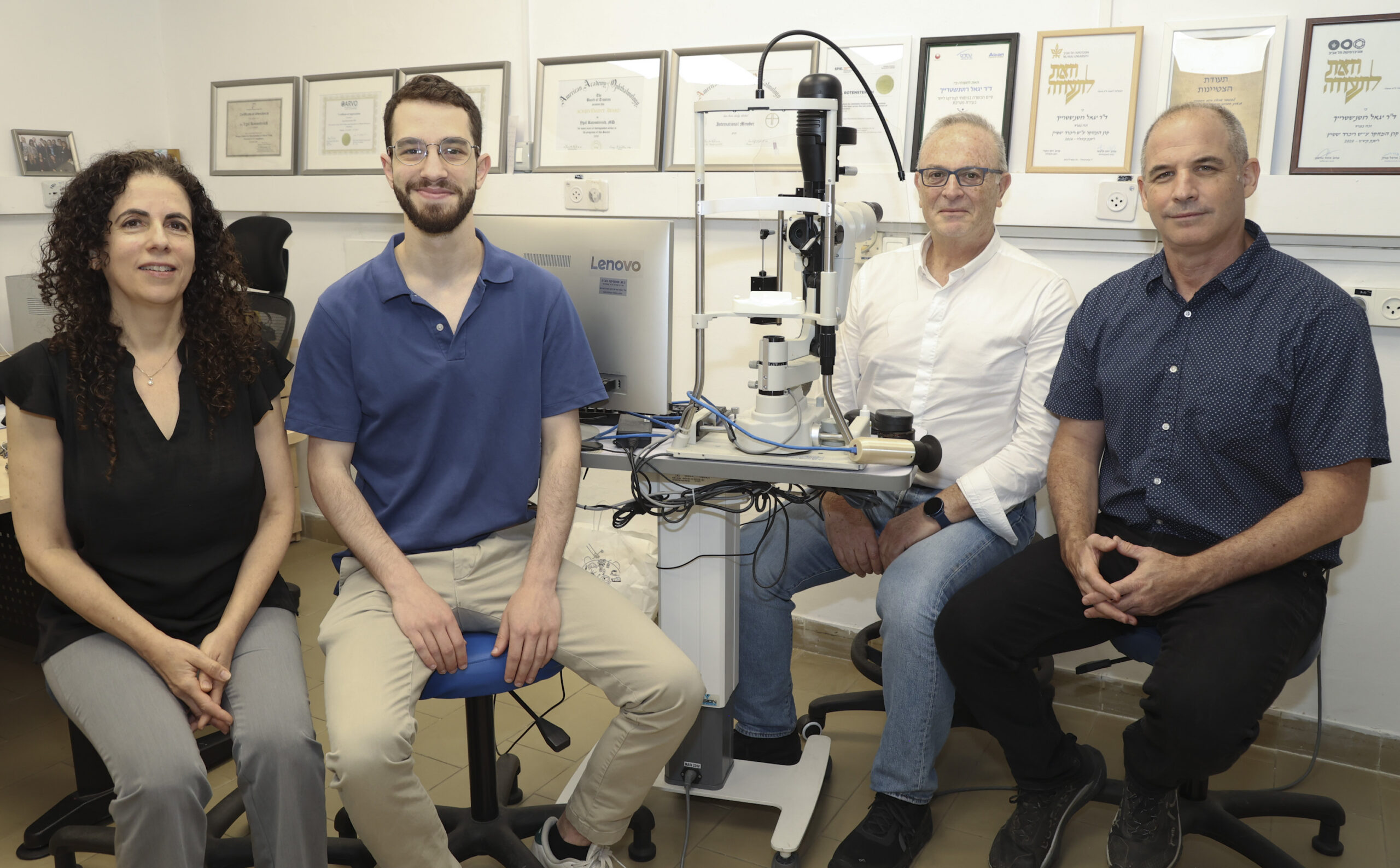
Left to Right: Dr. Ifat Sher-Rosenthal, Tamir Denis, Prof. Ygal Rotenstreich and Prof. Haim Suchowski Credit: Sheba Medical Center’s Photo Department
A new collaborative study by Tel Aviv University and Sheba Medical Center marks a significant advance toward non-invasive blood testing, one of the most significant unmet needs in the market.
Researchers have developed an artificial intelligence-based system capable of assessing hemoglobin levels and red blood cell counts using a short video recording of the blood vessels in the eye’s conjunctiva, the transparent membrane covering the white part of the eye, without the need for a needle prick or blood draw.
The study was conducted by Tamir Denis, a master’s graduate of Tel Aviv University, in collaboration with the research groups of Prof. Haim Suchowski of the School of Physics and Astronomy and Prof. Lior Wolf of the Blavatnik School of Computer Science and AI, together with researchers from Sheba Medical Center: Prof. Ygal Rotenstreich, Head of the Electrophysiology Clinic and Retinal Research Laboratory, and Dr. Ifat Sher-Rosenthal, Research Director, and Head of the Restorative Retinal Lab. The findings were published in the scientific journal npj Digital Medicine, part of the Nature portfolio of journals.
Blood tests are among the most commonly performed medical procedures worldwide, yet they still rely on invasive blood sampling and complex laboratory processing. Previous attempts focused on anatomical sites failed to demonstrate significant correlation. The researchers note that this new technology could eventually enable faster and more accessible testing, particularly in regions where access to healthcare services is limited. The study highlights that anemia is one of the most prevalent medical conditions in the world, affecting approximately 30% of the global population.
The study presents a technology called Video-to-Vessels, which converts high-magnification video recordings of the tiny blood vessels in the eye’s conjunctiva into a compact digital representation of vascular structure and blood-flow dynamics. This information is then fed into an artificial intelligence system trained to identify correlations between blood-flow characteristics and key blood markers, such as hemoglobin (Hb) levels and red blood cell (RBC) counts.
The study included 224 participants who underwent both standard blood tests and imaging of the conjunctiva using a 50x magnification RGB camera. Ten-second video recordings were collected from both eyes of each participant.
The study’s principal finding was that the system achieved a relatively high accuracy rate of 82.8% in detecting anemia. In addition, a strong correlation was found between the system’s predictions and laboratory results for both hemoglobin levels and red blood cell counts.
According to the researchers, one of the most notable examples of the system’s capabilities was its ability to detect subtle differences among extremely thin blood vessels. The study found that these vessels provided the most accurate information for predicting hemoglobin levels. The researchers explain that in very narrow vessels, blood cells move in single file, making it easier to identify blood-flow patterns and changes associated with hemoglobin concentration. Models trained exclusively on these small vessels achieved significantly better results than those based on larger vessels.
Another key finding was the importance of video processing. The researchers demonstrated that stabilizing eye movements and removing digital noise significantly improved the system’s performance. When these steps were omitted from the process, the predictive correlation declined by 38% for hemoglobin and by 19% for red blood cell counts.
The researchers emphasize that this remains a proof-of-concept study, and that broader, more diverse studies will be needed before the technology can be implemented in clinical practice. Nevertheless, they believe that it could eventually be developed into a compact handheld device to serve as a first-line screening tool in clinics, community healthcare settings, and even at home.
Tamir Denis explains: “One of the things that fascinated us most is the fact that in this region of the eye, not only can you see the blood vessels, but in some cases, you can actually observe the blood flow itself. This makes the conjunctiva a unique and highly compelling area for research. For us, this represents a new source of physiological information that, when combined with image processing and artificial intelligence, could entirely transform both the testing experience and access to it in the future. Our study is a first step in that direction. What excited me about this research from the very beginning was the sense that it has the potential to make a real difference in people’s lives, especially in places where access to medical infrastructure is limited.”
Prof. Ygal Rotenstreich concludes: “We view this study as a significant step toward the development of a new generation of non-invasive medical tests. The ability to extract information about a person’s blood profile using only a short video of blood vessels in the eye demonstrates the enormous potential of combining physics, optics, and artificial intelligence in medicine. Although this is still an early-stage study, the results are very encouraging and point to the future possibility of performing faster, simpler, and more accessible screening tests, even outside the hospital setting. We believe that technologies of this kind could eventually improve access to medical diagnosis and ease the burden on patients around the world.”
The research was supported in part by Israel’s Ministry of Innovation, Science and Technology.


